
Common Applications Of Pet-ct
PET-CT is a special type of nuclear medicine exam with an increasingly important range of diagnostic applications. It combines the key strengths of two imaging modalities—CT (computed tomography) and PET (positron emission tomography) to create 3D images with powerful diagnostic applications.
In a PET-CT scan, a small dose of radioactive material attached to sugar molecules—called a radiotracer—is administered to the patient. The radiotracer is taken up by cells of the body through the natural process of metabolizing the sugar.
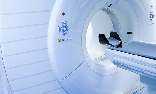
A special scanner that combines both PET and CT imaging technology is then used to create sets of image slices of tissue at different depths that combine the detailed, high-resolution computer-enhanced X-ray images of CT with color-coded PET images that show functional processes and changes as the radiotracer circulates through the body and is metabolized by cells.
The CT images allow physicians to examine tissues in fine detail to detect abnormalities and disease, while the PET images use color coding to represent different levels of metabolic activity. “Hot spots” in the PET images can indicate abnormalities such as higher metabolic activity in potentially cancerous tissue.
PET images can also provide information about other problems, such as impaired circulation in arteries with blockages.
PET-CT has an increasing role in diagnosis and treatment planning for a wide range of diseases and conditions. Among the most common purposes for ordering a PET-CT scan are:
-Cancer detection, diagnosis, staging, treatment planning, and monitoring during and after cancer treatment
-Assessing flow of blood to the heart muscle
-Determining the effects of a heart attack on the heart muscle
-Planning heart surgeries
-Evaluating abnormalities and disorders of the brain and central nervous system
843. 692. 0570 • 1303 Azalea Ct. • Ste. B • Myrtle Beach, SC 29577
© 2026 Carolina Radiology • Web Design by Three Ring Focus • Privacy / Terms / Employment